Using Reflectometry for a PPG Waveform
Abstract
The optical sensor is the most common type of biosensor. This application note provides an overview of the use of reflectometry for a pulse plethysmograph (PPG) waveform and describes the physical and physiological principals at work.
Introduction
Optical methods are among the most common approaches to biosensing for plants and animals. For example, remote s ensing equipment from satellites routinely use reflectance quality to determine the greenness and stresses of vegetation, and pulse oximeters utilizing pulse plethysmograph (PPG) sensors are used to collect vital-sign data during doctors’ visits. Indeed, optical sensing is rather versatile, as these application examples indicate. Light, whether coherent or non-coherent, interacts with matter as it passes through and becomes absorbed, reflected, scattered, dispersed, or otherwise altered. And scientists can examine the magnitude and shape of light pulses, their spectral contents, and polarization to derive information about the analytes in the media that the light pulses have traversed.
Monitoring Real-Time Blood Flow
A plethysmogram is a volumetric measurement. As blood flows, a cardiovascular pulse wave travels from the heart and propagates through the body, periodically distending the arteries and arterioles in the subcutaneous tissue. PPG uses a light to interrogate the piece of tissue, and the light received through the tissue corresponds with the variation of the blood volume.
Depending on the relative positions of the light source and the photodetector, two configurations are possible for PPG: transmissive absorption and reflection. In a transmissive configuration, the light source and the sensor are on directly opposite sides of the tissue. In a reflective arrangement, they could be on the same side. Reflective configurations take advantage of the light-scattering effect of body tissue.
In clinics and hospitals today, PPG is typically performed using a finger clip. However, as long as there’s easy access to tissue rich with blood vessels, it is possible to obtain a valid PPG signal in many other body locations. This is particularly true when using a reflective configuration. The forehead, the outer ear canal, the areas around the bicep or calve muscles, and even the area around a wrist are some examples of such body locations.
Optical Measurement Through Tissue
Visible to near-infrared (NIR) light can penetrate a depth of body tissues. Since light is absorbed by blood, melanin, fat, and water, penetration is limited. Within the range, the light is primarily scattered and, therefore, becomes diffused rapidly. Light-tissue interactions depend upon different tissue components and the wavelength of the light used. Given these variations, PPG signals can convey many pieces of information.
The backscattered light is modulated by pulsating arterial blood volumes, while absorption from other tissue components remains constant. In a simplified perspective, these result in an AC component in the photodetector output that is synchronous and proportional to the subject’s plethysmographic signal as well as a DC signal that is a function of the light source and the constant absorption by the tissue in the optical path. See Figure 1 for an example of backscattered light that a PPG photodetector receives, with the digitized photodetector output using three different light sources: red, green, and infrared. The signals have been processed to remove low-frequency noises.

Figure 1. Backscattered light received by the photodetector of a PPG.
The body absorbs light at different wavelengths to differing degrees. For example, many PPG devices include one or more green LEDs. Green light is readily absorbed by our bodies, so focusing on green light mitigates any contamination from reflected ambient light. However, because of the strong absorption, the depth of penetration is also limited, so it is suitable only to areas where blood perfusion is plentiful.
Hemoglobin also strongly absorbs green light, so it’s hard for this light to penetrate deeper into tissue. Medical implementations of PPG for pulse oximetry use NIR light sources. Red light penetrates deeply into the body, delivering a rich source of physiological signals.
Mitigating PPG Signal Artifacts
A PPG signal typically contains not only the backscattered light through the tissue but also artifacts introduced by ambient light and poor connectivity with the tissue. Various factors can affect the quality of a PPG signal, including the subject's skin structure, skin pigments, and even skin temperature. To mitigate the effects of some of these artifacts, while also conserving power, advanced PPG integrated circuits, such as the MAX30112, include specialized signal processing and sampling schemes.
For wearable-device designers, one of the biggest challenges involves addressing contamination from ambient light. Ambient light continuously changes, and indoor lights typically possess a flicker that tracks the powerline frequency (i.e., 50Hz or 60Hz, depending on location) that can obscure information carried in the AC component of a PPG signal.
Intermittently poor contact to the tissue and the photodetector can lead to motion artifacts. Such artifacts, in turn, can make it difficult to measure some slow physiological changes, particularly in wearable devices where the subject’s movement cannot be limited.
Another concern with PPG is that the detectable signal magnitude is affected by melanin concentration of the skin or skin pigments. Melanin, by design, attenuates the incident light wavelength. It is found in the epidermal layer, where there is no blood supply, so the target for a PPG device is always sub-epidermal. To overcome a weak signal due to dark pigmentation, designers can opt to use a stronger light source, or select a different light frequency, like NIR, where melanin is less absorbent.
Insights from PPG Are a Window into Cardiovascular Health
After artifacts have been removed and the PPG waveform is amplified through signal processing, advanced algorithms can be used to extract and interpret its features. See Figure 2 for a typical PPG waveform. A peak-peak interval, or the distance between two consecutive systolic peaks, represents a complete heart cycle.
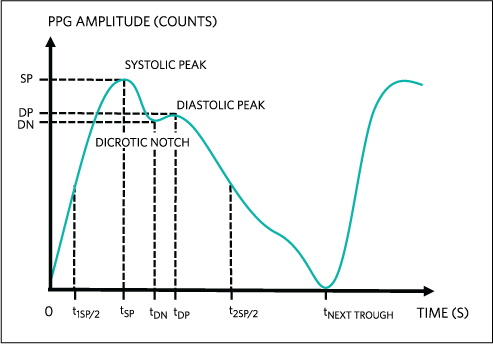
Figure 2. Typical PPG waveform.
The systolic amplitude (x) indicates the changes in blood volume caused by arterial blood flow and has been related to stroke volume, which could indicate vasoconstriction or vasodilation; it can also reflect cold sensitivity, reaction to anesthetics, blood loss, hypothermia, etc. Pulse width and pulse area also correlate with the systemic vascular resistance, which could reflect drug interaction of the patient or blood viscosity.
An indicator of aortic stiffness is the augmentation index, which is the ratio between the diastolic peak and systolic peak (y.x). The time delay between the systolic and diastolic peaks shortens with the subject’s age and, given the subject’s height, provides an indicator for large artery stiffness. Both of these indicators could inform a subject’s cardiovascular health.
We can also compare PPG signals from light sources at more than one wavelength. For instance, we can compare light absorption with a red and an infrared LED through the same tissue to determine the tissue’s level of oxygen saturation. Deoxyhemoglobin is more absorbent of red light than is oxyhemoglobin, while oxyhemoglobin can better absorb infrared. Blood oxygenation is, therefore, proportional to the ratio of reflected red and infrared lights.
PPG has either demonstrated success in, or has been proposed for use in, measuring parameters such as:
- Heart rate and heart rate variations
- Respiratory rate
- Blood oxygen saturation
- Body hydration
- Severity of venous reflux disease (varicose veins)
- Venous function
- Cold sensitivity
- Blood pressure
- Cardiac output
PPG a Practical Alternative for Heart-Rate Measurements
Easy to use and non-invasive, PPG has been used to measure a variety of physiological conditions. With advances in specialized PPG circuits, photoplethysmography is portable, low power, and relatively simple to implement. In clinical studies comparing PPG to electrocardiogram (ECG) for long-term heart rate variability (HRV) measurements, PPG has been shown to be a practical alternative to ECG. Even so, note that PPG can provide false readings if the photodetector cannot maintain good contact with the tissue (as a result of misinterpreted motion artifacts).
参考电路
1 Nakajima K, Tamura T, Miike H. Monitoring of heart and respiratory rates by photoplethysmography using a digital filtering technique. Med Eng Physics 1994; 18(5): 365-72.
2 Moore D, Maher T, Kingston V, Shanik G. Assessment of venous function using photoplethysmography. Ir J Med Sci 1982; 151(1): 308-12.
3 Lees T, Lambert D. Patterns of venous reflux in limbs with skin changes associated with chronic venous insufficiency. British Journal of Surgery. 1993;80(6):725-8.
4 Liu J, Yan B, Dai W, Ding X, Zhang Y, Zhou, N. Multi-wavelength photoplethysmography method for skin arterial pulse extraction. Biomedical Optics Express 7(10): 4313-4326
5 Weinschenk S, Beise R, Lorenz J. Heart rate variability (HRV) in deep breathing test and 5-min short-term recordings: agreement of ear photoplethysmography with ECG measurements, in 343 subjects. European Journal of Applied Physiology (2016) 116:1527-1535
A similar version of this application note appeared on EDN on October 11, 2017.